Uveitis: Don’t ignore
Uveitis may occur as a normal immune response to fight an infection inside the eye. Research suggests that there may be a link between black tattoo ink and uveitis. It is thought that skin tattoeing may trigger an immune response……
By Dr Shishir Narayan
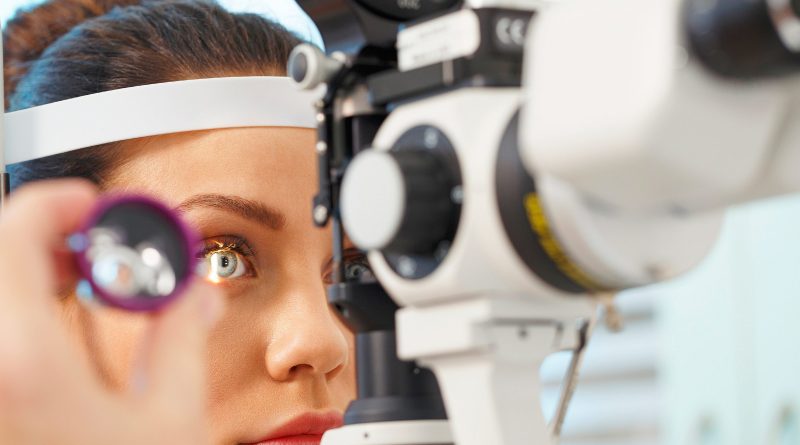
If you have eye redness, pain, blurry or lessened vision and sensitivity to light and floaters it is important to go see your eye doctor. You may have Uveitis which is basically eye inflammation and swelling that can destroy eye tissues. If you don’t take it serious it can lead to poor vision or blindness. Prompt diagnosis and treatment can help save your vision.
The word “uveitis” is used because the swelling most often affects the part of your eye called the uvea. An eye is made of layers. The uvea is the middle layer. It is right between the white part of your eye — called the sclera — and the inner layers of your eye
According to Dr Shishir Narayan, Senior Eye Specialist, Shroff Eye Hospital, New Delhi. Uveitis refers generally to a range of conditions that cause inflammation of the middle layer of the eye, the uvea, and surrounding tissues. It can be painful, the eye or eyes may be red, and vision may be cloudy.
An injury to the eye, a viral or bacterial infection, and some underlying diseases may cause uveitis. It can cause swelling and damage in the tissues of the eye. Untreated, it can lead to vision loss. It can affect one or both eyes.
The term uveitis is not only used to refer to an inflammation of the uvea, but to any part of the inside of the eye. It is not a single disease, and it has different causes. It is the fifth leading cause of vision loss in the United States, and so it has serious social and economic implications. It mainly affects people aged from 15 years to 40 years and above.
An uvea contains three important structures like The iris: That’s the colored circle at the front of your eye, The ciliary body: Its job is to help your lens focus and keep it healthy and The choroid: This is a group of blood vessels that give your retina the nutrients it needs.
There are different types of Uveitis like Anterior uveitis which is the most common. It affects the front of your eye, Intermediate : it uveitis affects your ciliary body and Posterior uveitis: it affects the back of your eye.If your entire uvea is inflamed, you have pan-uveitis.
What Causes It?
The exact cause of uveitis is often unclear, but some factors increase the chance of it happening.These include Juvenile arthritis, psoriasis and other autoimmune disorders, such as rheumatoid arthritis. Inflammatory disorders, such as Crohn’s disease, ulcerative colitis, AIDS/HIV and other diseases that weaken the immune system,
Uveitis may occur as a normal immune response to fight an infection inside the eye. Research suggests that there may be a link between black tattoo ink and uveitis. It is thought that skin tattoeing may trigger an immune response that affects both the eyes and the skin, in some people.
Types
There are different types of uveitis.
Anterior uveitis is also known as iritis, affects the colored part of the eye, the iris. Iridocyclitis is similar, but it include inflammation of the ciliary body. Intermediate uveitis can be vitritis or pars planitis. Vitritis is an inflammation of the jelly-like part of the eye, the vitreous cavity. An inflammation of the pars plana is called pars planitis. Posterior uveitis is an inflammation of the retina and choroid. Posterior refers to the back of the eye. Pan-uveitis is an inflammation in all layers of the uvea.
Signs and symptoms of Uveitis
•General vision problems, including blurred or cloudy vision
•Floaters, spots in the eye that look like tiny rods or chains of transparent bubbles floating around in the field of vision
•Eye pain and redness
•Photophobia, an abnormal sensitivity to light
•Headaches
•A small pupil
•Alteration of the color of the iris
Symptoms can appear on gradually or rapidly.
Diagnosis
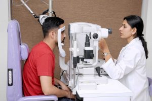
An ophthalmologist, or eye specialist, will ask about signs, symptoms, and general medical history. A doctor will look at symptoms and check for underlying conditions.It is important to know whether the uveitis is caused by an infectious process or an underlying disease.If another condition appears to underlie the uveitis, the ophthalmologist may refer the patient to a specialist to make sure that condition receives proper treatment.The ophthalmologist will look at the eye with a special slit lamp. When the light hits the inside of the eye, the doctor can determine whether that area is clear or foggy.
If there is inflammation in the iris, patients may feel some pain when the pupil contracts, which is when light hits it.If uveitis is present, white blood cells and protein in the eye fluid can be seen through the microscope.
Treatment
A patient with uveitis who receives prompt and appropriate treatment will usually recover. Without treatment, there is a risk of cataracts, glaucoma, band keratopathy, retinal edema, and permanent vision loss.
Antibiotics or antiviral medication will be used if there is an infection. Corticosteroid medications are sometimes given as well, in the form of eye drops (prednisolone acetate), tablets, or as an injection into the eye. Steroids are effective in treating inflammation. Before giving corticosteroids, it is important rule out corneal ulcers by using a florescence dye test.
Immunosuppressants might be recommended if symptoms are very severe and there is a risk of vision loss, or if the patient has not responded well to other therapies.
Mydriatic eye drops, such as atropine or cyclopentolate, dilate the pupil and help the eye to heal. It also helps with eye pain and stops the pupil from sticking to the lens. There may be blurred vision and unusual sensitivity to light, known as photophobia.
Complications
With prompt and proper treatment and close monitoring, the chances of complications are significantly reduced. If they do occur, they may include Glaucoma, Cataracts, Macular edema, Scar tissue, Retinal detachment, or detached retina and Vision loss.
(The author is Senior Eye Specialist, Shrooff Eye Centre, New Delhi)
How is Uveitis Treated?
Uveitis treatments primarily try to eliminate inflammation, alleviate pain, prevent further tissue damage, and restore any loss of vision. Treatments depend on the type of uveitis a patient displays. Some, such as using corticosteroid eye drops and injections around the eye or inside the eye, may exclusively target the eye whereas other treatments, such immunosuppressive agents taken by mouth, may be used when the disease is occurring in both eyes, particularly in the back of both eyes.
An eye care professional will usually prescribe steroidal anti-inflammatory medication that can be taken as eye drops, swallowed as a pill, injected around or into the eye, infused into the blood intravenously, or, released into the eye via a capsule that is surgically implanted inside the eye. Long-term steroid use may produce side effects such as stomach ulcers, osteoporosis (bone thinning), diabetes, cataracts, glaucoma, cardiovascular disease, weight gain, fluid retention, and Cushing’s syndrome. Usually other agents are started if it appears that patients need moderate or high doses of oral steroids for more than 3 months.
Other immunosuppressive agents that are commonly used include medications such as methotrexate, mycophenolate, azathioprine, and cyclosporine. These treatments require regular blood tests to monitor for possible side effects. In some cases, biologic response modifiers (BRM), or biologics, such as, adalimumab, infliximab, daclizumab, abatacept, and rituximab are used. These drugs target specific elements of the immune system. Some of these drugs may increase the risk of having cancer.
Anterior Uveitis Treatments
Anterior uveitis may be treated by:
•Taking eye drops that dilate the pupil to prevent muscle spasms in the iris and ciliary body (see diagram).
•Taking eye drops containing steroids, such as prednisone, to reduce inflammation.
Intermediate, Posterior, and Pan-Uveitis Treatments
Intermediate, posterior, and pan-uveitis are often treated with injections around the eye, medications given by mouth, or, in some instances, time-release capsules that are surgically implanted inside the eye. Other immunosuppressive agents may be given. A doctor must make sure a patient is not fighting an infection before proceeding with these therapies.
A recent NEI-funded study, called the Multicenter Uveitis Treatment Trial (MUST), compared the safety and effectiveness of conventional treatment for these forms of uveitis, which suppresses a patient’s entire immune system, with a new local treatment that exclusively suppressed inflammation in the affected eye. Conventionally-treated patients were initially given high doses of prednisone, a corticosteroid medication, for 1 to 4 weeks which were then reduced gradually to low doses whereas locally-treated patients had a capsule that slowly released fluocinolone, another corticosteroid medication, surgically inserted in their affected eyes. Both treatments improved vision to a similar degree, with patients gaining almost one line on an eye chart. Conventional treatment produced few side effects. In contrast, the implant produced more eye problems, such as abnormally high eye pressure, glaucoma, and cataracts. Although both treatments decreased inflammation in the eye, the implant did so faster and to a greater degree. Nevertheless, visual improvements were similar to those of patients given conventional treatment.
The National Eye Institute conducts and supports a number of studies investigating uveitis, including:Testing new methods for treating uveitis. Investigating the possible causes of uveitis.Learning who is most susceptible to the disease. This research should provide better ways to detect, treat, and prevent uveitis.
Anterior Uveitis: Most commonest form of intraocular inflammation
Research is still ongoing to find out who is most likely to develop uveitis, the possible causes, and new ways of treating it. Anterior uveitis is the commonest form of intraocular inflammation with a varying incidence in the general population of various countries around the world. The potential severe consequences of recurrent or untreated anterior uveitis are probably underestimated.
Anterior uveitis involves inflammation of the iris alone (iritis), anterior part of ciliary body (anterior cyclitis) or both structures (iridocyclitis). It is commoner than posterior segment inflammation and is generally less sight-threatening and less serious, especially if treated early. Anterior uveitis normally causes reduction in the vision during the acute stage but it is the sequelae of anterior uveitis which can have long-lasting impact.
Detailed medlars search was carried out to review the articles and case reports on anterior uveitis. The methods used to collect/select evidence were hand-searches of published literature (primary sources) and searches of electronic databases. The current guidelines for diagnosis and management of anterior uveitis are based on the literature search using the National Library of Medicine’s Medline database and the Vision Net database.
The uveitis was classified in different ways. Classification based on the duration of uveitis was based on Standardization of Uveitis Nomenclature (SUN) criteria in which anterior uveitis was classified as limited (less than or equal to three months duration) and persistent (more than three months). Based on the course of uveitis, anterior uveitis was classified as acute anterior uveitis with episodes of sudden onset and limited duration, recurrent anterior uveitis with repeated episodes separated by periods of inactivity without treatment ≥ three months in duration, and chronic uveitis which persists and relapses in less than three months after discontinuing treatment.
Based on etiology anterior uveitis was classified as infectious (such as viral, bacterial, fungal or protozoal), autoimmune with only ocular involvement or with systemic disease association or presenting as masquerade syndrome. Anterior uveitis with other etiologies can be post-traumatic, post-surgical, lens-induced and drug-induced. Pathologically anterior uveitis was classified as granulomatous or non-granulomatous based on the nature of keratic precipitates.
Anterior uveitis can present with acute, chronic or recurrent attacks. Anterior uveitis is the commonest type of intraocular inflammation and commonly presents as unilateral presentation with pain or photophobia, circumlimbal redness and anterior chamber cells and flare. Patients with anterior uveitis usually complain of pain, redness, blurred vision, and photophobia, watering.
Most of the patients would have had repeated attacks and would have sought consultation with multiple ophthalmologists and would have used topical and/or systemic medications on and off. Blurring of vision, which is perhaps the commonest symptom, is caused by turbidity of the aqueous. Photophobia is commonly due to ciliary muscle spasm but anterior chamber cellular infiltration, corneal epithelial edema and pupillary muscle involvement can also contribute. Varying degree of pain seen in anterior uveitis can be attributed to ciliary muscle spasm. It is usually a dull aching type of pain or a throbbing sensation. Severe pain can be associated with raised intraocular pressures. The common clinical signs with which a patient of anterior uveitis can present are:
Nil to varying degrees of lid edema may be a presenting sign in patients with anterior uveitis. Circumcorneal congestion may be seen due to enlargement of the episcleral vessels in the region of the ciliary body. Keratic precipitates (KPs) are cellular deposits on the corneal endothelium. Fine KPs are presumed to be of non-granulomatous allergic type of inflammation whereas large and mutton fat KPs are associated with granulomatous inflammation. Colored or pigmented KPs suggest prior episodes of anterior uveitis. Microscopically, KPs are accumulation of lympho-plasmocytic inflammatory cells, with epitheloid cells seen additionally in granulomatous KPs.
Aqueous cells and flare are due to cellular infiltration and protein exudation into the anterior chamber. Aqueous cells are an early and definite sign of active inflammation. The translucence of the aqueous due to its high albumin content is called aqueous flare. It is an indefinite sign of active inflammation. Examination of the anterior chamber involves observing with high magnification while directing a small, intense beam obliquely through the aqueous, following relative dark adaptation. Anterior chamber cells and/or flare are visible owing to the Tyndall effect of the bright beam. The grading of cellular reaction in the anterior chamber helps in the assessment of the severity of anterior uveitis [Table 1]. Grading is useful in determining the patients’ response to therapy as well as long-term monitoring. Miosis can be due to reflex spasm of the sphincter or due to vascular distension of the iris vessels. Iris nodules are accumulations of leukocytes lying on the anterior iris; Koeppe’s nodules are seen at the pupillary margin whereas Busaca’s nodules are present on the anterior iris stroma.
The nodules on the surface of the iris need to be differentiated from infected nodules. Posterior synechiae are the adhesions between the anterior lens surface and the iris; posterior synechiae extending for 360° are called seclusio pupillae while occlusio pupillae refer to a membrane obscuring the lens surface; anterior chamber can show fibrinous reaction hypopyon, pupillary membrane with hypopyon and hyphema. Iris atrophy is associated with chronic iridocyclitis and occurs due to ischemia. Neovascularization can occur on the iris stroma or in the anterior chamber angle, which may lead eventually to neovascular glaucoma. Anterior vitreous cells are far exceeded by aqueous cells in iritis, whereas in iridocyclitis with intermediate uveitis the cells are distributed equally between the two compartments. Complicated cataract occurs due to thickened lens capsule due to posterior synechiae or altered membrane permeability.
Inflammation can result in either increased or decreased intraocular pressure. Acute attack of anterior uveitis with severe anterior chamber inflammation can lead to increase in intraocular pressure and is most commonly seen in viral keratouveitis or Posner Schlosman syndrome. However, idiopathic anterior uveitis can also present with raised intraocular pressure. Fuchs’ heterochromic iridiocyclitis is known to be associated with intractable open-angle glaucoma in late stages and the patients need to be counselled about the same. Severe inflammation of the ciliary body may lead to decreased aqueous production and subsequent fall in intraocular pressure may be a result of the inflammation itself, sequelae of inflammation, or because of the steroid treatment. Presence of cyclitic membrane over ciliary body in cases with chronic or recurrent intermediate uveitis with spillover anterior uveitis also leads to severe hypotony. In active inflammation, raised intraocular pressure can be due to associated trabeculitis or can be due to secondary angle closure. Though associated trabeculitis is not typical of any specific anterior uveitis entity it can be commonly seen in cases with viral keratouveitis. Gonioscopy would reveal gonio-synechiae or neovascularization in angles, and the angles could be open or closed angles depending on the stage of uveitis . The following features should be looked on fundus examination such as disc edema and hyperemia, vascular sheathing, perivascular exudates, cystoid macular edema, retinitis, chorodial infiltrates, retinal detachment, pars plana exudates or snow banking. Presence of positive findings on posterior segment examination indicates anterior uveitis as a part of panuveitis (e.g. sarcoidosis, tuberculosis, Vogt Koyanagi Harada Disease, sympathetic ophthalmia) or as spillover uveitis in cases with intermediate uveitis..
The treatment of uveitis
The treatment of uveitis is undergoing significant change as a result of the development of new therapeutic approaches, of which the biologic agents form a major part. These targeted therapies have shown great promise for the treatment of refractory disease and some have now undergone systematic evaluation through prospective clinical trials, unlike many of their predecess or drugs.
Uveitis is one of the leading causes of blindness worldwide. Noninfectious uveitis may be associated with other systemic conditions, like human leukocyte antigen B27-related spondyloarthropathies, inflammatory bowel disease, juvenile idiopathic arthritis, Behçet’s disease, and sarcoidosis. Conventional therapy with corticosteroids and immunosuppressive agents (such as methotrexate, azathioprine, mycophenolate mofetil, and cyclosporine) may not be sufficient to control ocular inflammation or prevent non-ophthalmic complications in refractory patients. Off-label use of biologic response modifiers has been studied as primary and secondary therapeutic agents. They are very useful when conventional immunosuppressive therapy has failed or has been poorly tolerated, or to treat concomitant ophthalmic and systemic inflammation that might benefit from these medications.
Biologic therapy, primarily infliximab, and adalimumab, have been shown to be rapidly effective for the treatment of various subtypes of refractory uveitis and retinal vasculitis. Other agents like golimumab, abatacept, canakinumab, gevokizumab, tocilizumab, and alemtuzumab may have great future promise for the treatment of uveitis. It has been shown that with proper monitoring, biologic therapy can significantly improve quality of life in patients with uveitis, particularly those with concurrent systemic symptoms.
 According to Dr Mahipal S Sachdeva, Chairman and Medical Director, Centre for Sight, New Delhi, however, given high cost as well as the limited long-term safety data, we do not routinely recommend biologics as first-line therapy for noninfectious uveitis in most patients. These agents should be used with caution by experienced clinicians. The present work aims to provide a broad and updated review of the current and in-development systemic biologic agents for the treatment of noninfectious uveitis.
The term “uvea” comes from the Latin word for grape. The eye includes three layers. The middle layer, or uvea, encompasses the iris, ciliary body, and choroid. Inflammation of the uvea is termed uveitis, but it is usually diagnosed on the basis of inflammation in adjacent structures which include the anterior chamber, the vitreous humor, or the retina. Inflammation in the uvea can be due to infections, masquerades such as B-cell lymphoma, or immune-mediated diseases. The latter can be a systemic disease such as sarcoidosis or a disease confined to the eye such as sympathetic ophthalmia. Anatomic classification of uveitis is extremely useful, since the differential diagnosis is distinct for anterior, intermediate (involving the vitreous humor), posterior (involving the retina or choroid), and panuveitis.1
 Dr Rajesh Ranjan, Senior Eye Specialist, Eye 7 Hospital, Indiapuram (Ghaziabad), said, “Uveitis is the third leading cause of blindness in the developed countries. The annual incidence is estimated between 17 and 52 per 100,000 persons, and the prevalence is 38–714 per 100,000 persons.2 The incidence and prevalence vary among different geographic locations worldwide. Males and females are generally equally affected overall, but sex preponderance may be observed in some uveitis groups, such as male predominance in human leukocyte antigen (HLA)-B27-associated uveitis and female preponderance in juvenile idiopathic arthritis (JIA)-related uveitis. Uveitis may occur at any age, but most commonly affects the working population aged between 20 and 59 years. Childhood uveitis is relatively less common, but may cause long-term severe visual loss.2 Therefore, the burden of this sight-threatening condition is very significant.”
 Dr. Prof. L.D. Sota, MS – Ophthalmology, Max Super Speciality Hospital Vaishali. said, “The most common symptoms of uveitis are decreased vision, eye pain, redness, light sensitivity, and floaters. The redness and eye pain are generally seen in eyes with acute anterior inflammation, but may not be prominent in chronically inflamed eyes or those in which the inflammation is confined only to the posterior segment.”
Uveitis is typically an immune-mediated condition, which involves chemical mediators resulting in vascular dilation (conjunctival injection), increased vascular permeability (aqueous flare), and chemotaxis of inflammatory cells into the eye (aqueous and vitreous cellular response). With variable chronicity and severity, uveitis may be complicated by cataract, glaucoma, band keratopathy, hyphema, vitreous hemorrhage, cystoid macular edema (CME), retinal detachment, retinal ischemia, optic atrophy, chronic eye pain, and blindness.
Uveitis can be caused by infectious and noninfectious etiologies. Causative infectious origins may include bacteria, viruses, fungi, and parasites. The precise diagnosis is crucially important to establish an appropriate therapy. Specific antimicrobial treatment is typically required for infectious uveitis. In rare occasions, neoplastic diseases (eg, lymphoma) may masquerade as ocular inflammation, and an appropriate diagnosis is needed for proper management.
For noninfectious uveitis, excluding masquerade neoplasms, the control of inflammation is the key to treatment success. We generally use a stepladder approach; the treatment includes local corticosteroids, systemic corticosteroids, and systemic immune modulators, often sequentially starting with topical therapy. Noninfectious uveitides are often associated with other systemic conditions, such as HLA-B27-related spondyloarthropathies, inflammatory bowel disease (IBD), JIA, Behçet’s disease (BD), and sarcoidosis. The treatment of systemic symptoms may also improve ocular inflammation.