
Clouds in the vision
Cataract is no longer an inevitable fact of aging, but requires utmost farsightedness in its prevention, treatment, and post-operative care, whether in the case of adults or children
By Abhinav/Abhigyan
If you have even the slightest symptoms of cataract, you must see an eye specialist without losing any time.There is no conclusive evidence that shows why the eye’s lens changes as people advance in years, leading to formation of cataract. But researchers worldwide have identified factors besides advancing age that may cause cataract or are associated with cataract development.
Cataract can be termed as clouding of the eye’s natural lens, which lies behind the iris and the pupil. It is the most common cause of vision loss in people who have crossed the age of 40. Worryingly,cataract is the principal cause of blindness in the world. In fact, the cases of cataract worldwide outnumber the cases of glaucoma, macular degeneration and diabetic retinopathy combined.
Types of cataracts
1 Subcapsular cataract occurs at the back of the lens. People with diabetes or those taking high doses of steroid medications have a greater risk of developing a subcapsular cataract.
2 A nuclear cataract forms deep in the central zone (nucleus) of the lens. Nuclear cataracts usually are associated with aging.
3 A cortical cataract is characterized by white, wedge-like opacities that start in the periphery of the lens and work their way to the centre in a spoke-like fashion. This type of cataract occurs in the lens cortex, which is the part of the lens that surrounds the central nucleus.
Cataract Symptoms and Signs
A cataract starts at a small level and at first does not have much effect on your vision. You may notice that your vision is blurred a little, like looking through a cloudy piece of glass or viewing an impressionist painting. A cataract may make light from the sun or a lamp may seem too bright or glaring. Or you may notice when you drive at night that the oncoming headlights cause more glare than before. Colours may not appear as bright as they once did.
The type of cataract you have will determinethe symptoms you experience and the frequency of their recurrence. When a nuclear cataract first develops, it can bring about a temporary improvement in your near vision, called “second sight.” Unfortunately, the improved vision is short-lived and will disappear as the cataract worsens. On the other hand, a subcapsular cataract may not produce any symptoms until it’s well-developed.
What Causes Cataract?
The lens inside the eye works much like a camera lens, focusing light onto the retina for clear vision. It also adjusts the eye’s focus, letting us see things clearly both up close and far away. The lens is mostly made of water and protein. The protein is arranged in a precise way that keeps the lens clear and allows light to pass through it.But as we age, some of the protein may clump together and start to cloud a small area of the lens. This is a cataract, and over time, it may grow larger and cloud more of the lens, making it harder to see.
Besides advancing age, cataract risk factors include:
• Ultraviolet radiation from sunlight and other sources
• Diabetes
• Hypertension
• Obesity
• Smoking
• Prolonged use of corticosteroid medications
• Statin medicines used to reduce cholesterol
• Previous eye injury or inflammation
• Previous eye surgery
• Hormone replacement therapy
• Significant alcohol consumption
• High myopia
• Family history
One theory of cataract formation that’s gaining acceptance is that many cataracts are caused by oxidative changes in the human lens. This is supported by nutrition studies that show fruits and vegetables high in antioxidants may help prevent certain types of cataracts.
Cataract Prevention
Though it is debatable whether cataracts can be prevented, a number of studies suggest certain nutrients and nutritional supplements may reduce your risk of cataracts. One extensive, 10-year study of female health professionals found that higher dietary intakes of vitamin E and the carotenoids lutein and zeaxanthin from food and supplements were associated with significantly decreased risks of cataract. Good food sources of vitamin E include sunflower seeds, almonds and spinach. Good sources of lutein and zeaxanthininclude spinach, kale and other green, leafy vegetables. Other studies have shown that antioxidant vitamins such as vitamin C and foods containing omega-3 fatty acids may reduce cataract risk.
Cataract Treatment
When symptoms begin to appear, you may be able to improve your vision for a while using new glasses, strong bifocals, magnification, appropriate lighting or other visual aids. Think about surgery when your cataracts have progressed enough to seriously impair your vision and affect your daily life.
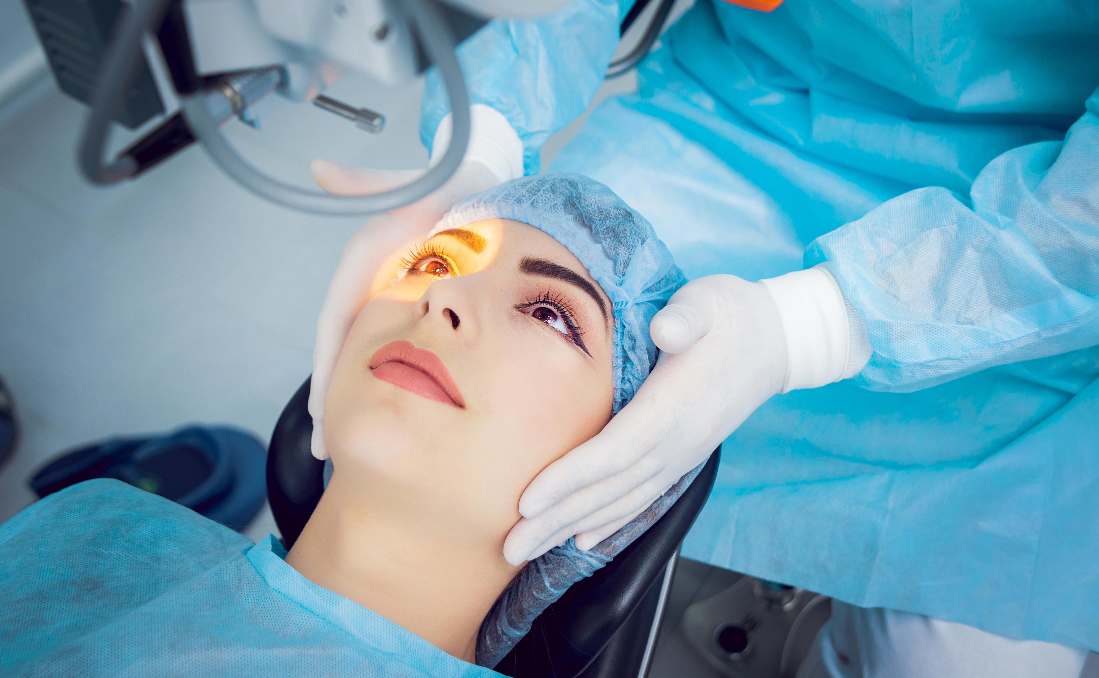
Many people consider poor vision an inevitable fact of aging, but cataract surgery is a simple, relatively painless procedure to regain vision.
Cataract surgery is very successful in restoring vision. Nine out of 10 people who have cataract surgery regain very good vision, somewhere between 20/20 and 20/40.
During surgery, the surgeon will remove your clouded lens and in most cases replace it with a clear, plastic intraocular lens (IOL). New IOLs are being developed all the time to make the surgery less complicated for surgeons and the lenses more helpful to patients. Presbyopia-correcting IOLs potentially help you see at all distances, not just one. Another new type of IOL blocks both ultraviolet and blue light rays, which research indicates may damage the retina.
Eyewear After Cataract Surgery
In most cases, unless you choose presbyopia-correcting IOLs, you will still need reading glasses after cataract surgery. You may also need progressive lenses to correct mild residual refractive errors as well as presbyopia.
For the best vision and comfort possible with glasses prescribed after cataract surgery, ask your optician to explain the benefits of anti-reflective coating and photochromic lenses.
Paediatric cataract
As per latest report released by World Health Organization (WHO), cataract is the leading cause of preventable blindness. The statistics show the cataract is the leading cause of blindness even during childhood. Diagnosis of paediatric cataract is not difficult; however management is more complex than cataract in the adult.
The surgical expertise needed is at a higher level. Experience is needed in terms of decision making regarding timing of the surgery, spacing of cataract surgery between two eyes, whether to go for an intra ocular lens implantation or not. One needs to be well versed with the primary posterior capsulotomy to avoid posterior capsular opacification and consequent amblyopia. Postoperative care needs to be more aggressive in terms of treatment of inflammation and visual rehabilitation. One needs to keep setting of amblyopia in mind, in these children.
If needed amblyopia treatment should to be instituted very early in the post-operative period to have optimal visual recoveries. However, the screening of children for white reflex should be taken up along the lines of a public campaign. The impact of a child going blind is enormous as it corresponds to the loss of number of man years of productivity. The Academic and Research committee has brought out a CME programme on pediatric cataract with an intention to increase awareness among ophthalmologists. This CME gives an excellent overview of clinical features, diagnosis and management of paediatric cataract. It can serve as good guide to approach a child with a paediatric cataract.
In this CME, the authors have demonstrated good surgical approach in a child with paediatric cataract and IOL power calculations in this age group. They also discussed appropriately the risks of amblyopia and its management especially in unilateral and bilateral cataracts and their care in the post-operative period. The reviewers have done a good job in pointing out the relevant lacunae, so that the CME series can be as useful as possible to all ophthalmologists.
Paediatric cataract is one of the major causes of preventable childhood blindness, affecting approximately 200,000 children worldwide, with an estimated prevalence ranging from three to six per 10,000 live births.1 to 3 paediatric cataracts may be congenital if present within the first year of life, developmental if present after infancy, or traumatic. Early diagnosis and treatment are of crucial importance to prevent the development of irreversible stimulus-deprivation amblyopia. The management of paediatric cataract should be customized depending upon the age of onset, laterality, morphology of the cataract, and other associated ocular and systemic comorbidities.
Recent advances in surgical techniques, intraocular lens (IOL) composition and designs, increased understanding about the neurobiology of visual development, and early postoperative use of contact lenses for optical rehabilitation have contributed to improved outcomes after paediatric cataract surgery. Furthermore, early diagnosis can be achieved by genetic counseling and testing in cases of hereditary cataracts.
However, certain issues specific to paediatric eyes, such as increased postoperative inflammation, axial growth after cataract extraction, implant-power calculation, secondary glaucoma, posterior-capsule opacification (PCO), and amblyopia management, are still major obstacles to achieving good visual outcomes in childhood cataract surgery.
The evaluation of a child with a cataract begins with a detailed history including family history; a prenatal history including maternal drug use and febrile illnesses with rash; and birth history, especially birth weight, since low birth weight may be associated with idiopathic bilateral congenital cataracts.A developmental history should be carefully assessed, and if required, review should be sought to exclude metabolic or systemic related etiologies. A history of the onset of the lenticular opacities, laterality, and progression is also important. Unilateral cataracts are usually isolated, but they are most commonly found to be associated with persistent foetal vasculature (PFV)also, other ocular abnormalities, such as lenticonus/lentiglobus may be associated.
A detailed ocular examination is carried out either in the office or in the operating room. This should include slit-lamp biomicroscopy to assess the size, location, density of lenticular opacity, capsular changes, such as preexistent posterior capsular defects, and other associated anterior-segment developmental anomalies. In addition, measurement of intraocular pressures and corneal diameters are performed.
Fundus examination in partial cataracts and ultrasound examination in total cataracts may reveal posterior-segment abnormalities that may affect the visual outcome. Ultrasound biomicroscopy can be informative in children with anterior-segment developmental anomalies and PFV, and also in the assessment of posterior capsular support while considering secondary IOL implantation. In children under 12 months of age, it is sometimes possible to examine them after they have been fed milk.
A cataract is any light scattering opacity of the lens. It is estimated that congenital cataracts are responsible for 5% to 20% of blindness in children worldwide. Incidence varies from country to country. One retrospective study of the prevalence of infantile cataracts in the US showed a rate of 3-4 visually significant cataracts per 10,000 live births. This is a similar rate to a UK study which showed 3.18 per 10,000. These numbers underestimate the total number since they do not take into consideration visually insignificant cataracts.
Cataracts may be unilateral or bilateral and can vary widely in size, morphology and degree of opacification from a small white dot on the anterior capsule to total opacification of the lens. Consequently, the effect on vision, course of treatment and prognosis may also be widely variable.
The causes of infantile cataracts have been the source of much speculation and research. Making a distinction between unilateral and bilateral cataracts may be useful when considering etiology.
The majority of bilateral congenital or infantile cataracts not associated with a syndrome have no identifiable cause. Genetic mutation is likely the most common cause. Over fifteen genes involved in cataract formation have been identified, and the inheritance is most often autosomal dominant although it can be X-linked or autosomal recessive. Within the same pedigree, there can be considerable morphologic variation.
Systemic associations include metabolic disorders such as galactosemia, Wilson disease, hypocalcemia and diabetes. Cataracts may be a part of a number of syndromes, the most common being trisomy 21. Intrauterine infections including rubella, herpes simplex, toxoplasmosis, varicella and syphilis are another causes.
In contrast, most unilateral cataracts are not inherited or associated with a systemic disease and are of unknown etiology although they do not rule out the possibility of an associated systemic disease. They are usually the result of local dysgenesis and may be associated with other ocular dysgenesis such as persistent fetal vasculature (PFV), posterior lenticonus or lentiglobus.
Trauma is a known cause of paediatric cataracts. If there is no known history of trauma to explain an acquired cataract in this age group, investigation must be considered in children who present with other signs suggestive of child abuse.
Regardless of the etiology, prompt treatment of visually significant cataracts is necessary to allow proper development of vision.
In many cases of congenital cataracts, there is a family history. History of prenatal and pregnancy history can also provide clues.Cataracts present as an opacity in the lens which run a spectrum from easily visible in the undilated state and apparent to the parents or pediatrician, to much more subtle changes requiring dilation and careful examination with a slit lamp. The red reflex is an extremely useful part of the exam giving an estimate of size and location within the visual axis, even in an uncooperative child.
Cataracts are classified according to their morphological appearance and location; however, making the diagnosis of a specific type of cataract can be difficult if it spreads to involve multiple layers, obscuring the original opacity. Cataracts may be a part of another disease or syndrome, and are sometimes the initial finding that leads to the diagnosis. A cataract may be accompanied by additional noticeable ocular abnormalities such as microcornea, megalocornea, coloboma of the iris, aniridia, and zonular dehiscence.
Symptoms
Often an infant with mild cataracts appears asymptomatic, delaying the diagnosis for years. At other times, lack of reaction to light, strabismus, a failure to notice toys and faces or an apparent delay in development become the cause of concern. Mild cataracts may cause photophobia only in bright lights. Dense cataracts also may be discovered if they lead to the development of sensory nystagmus.
Clinical diagnosis
For unilateral cataracts in an otherwise healthy child, an extensive workup is not necessary. The most critical part of the workup is a thorough ophthalmologic exam including slit lamp examination of both eyes, checking intraocular pressure, and an ultrasound of the posterior pole if not visible. If the exam reveals the classic appearance of a specific diagnosis such as PFV or posterior lenticonus, no further evaluation is necessary.
The first step in the workup of bilateral cataracts should be a family history including examination of family members. If there is a clear autosomal dominant pattern and the child is healthy, further evaluation is not necessary. In cases without clear family history, a thorough pediatric and developmental exam should be performed. Recommended lab workup includes TORCH titers, VDRL, serum calcium and phosphorus levels and urine for reducing substance. Additional systemic workup should be done in coordination with the paediatrician. Dysmorphic features may suggest the need for involvement of a geneticist.
Not all pediatric cataracts require surgery. A small, partial or paracentral cataract can be managed by observation. Pharmacologic pupillary dilation with phenylephrine or tropicamide can be helpful. Dilation with atropine should be avoided as it is amblyogenic. Part-time occlusion may be necessary in unilateral or asymmetric cases that develop or are at risk for amblyopia. These techniques may at least delay the need for surgery until a point when eye growth has stabilized and an IOL can be implanted with less refractive uncertainty. Because of the unpredictability in the progression of partial cataracts, these patients should be carefully monitored and if significant amblyopia develops that is unresponsive to treatment, surgical intervention should be performed.
Decision about surgery depends upon age of patient at presentation, extent of opacity and associated conditions.
If child is less than1 year sequence should be
Lensectomy pars plana or limbal leaving anterior capsular rim
Optic rehabilitation using aphakic glasses/contact lenses
Secondary implant in sulcus
Amblyopia and strabismus management
If child is 1 to 5
Primary surgery with anterior approach with primary posterior capsulotomy and anterior vitrectomy
If child is greater than 5
May not need primary capsulotomy
Surgery
If the cataract(s) are felt to be visually significant, surgical intervention is the only option. The timing of surgery is critical for visual development. Most investigators recommend surgery within the first two months of life. There has been evidence to suggest that before one month of age, the risk of aphakic glaucoma is increased. In cases of bilateral cataracts, it may be advantageous to perform surgery on both eyes in the same intervention to allow for simultaneous initiation of visual rehabilitation as well as reducing exposure to general anesthesia. In this setting, treating each eye as a separate sterile procedure may reduce infection risk.
Removal of the lens can be approached through the limbus or the pars plana. The limbal approach has the advantage of maintaining the posterior capsule to facilitate posterior chamber intraocular lens (IOL) implantation if desired.
Several options exist to open the anterior capsule in paediatric cataracts. The ideal anterior capsulectomy technique is one that results in low incidence of radial tears and is easily performed. In cases of dense cataract, dye can be used to stain the anterior capsule, making this step easier and safer. A manual continuous curvilinear capsulorhexis (CCC), which is the preferred method in adult eyes, can be difficult in paediatric cases due to the elasticity of the paediatric capsule. However, when it can be controlled and completed, it creates an edge, which has a low incidence of radial tears. A 23 G vitreous end gripping forceps using push pull technique is very effective.
A mechanized circular anterior capsulectomy, known as vitrectorhexis has been proven to be a very good, safe alternative if the CCC is not possible. The vitrector tip is placed through a stab incision at the limbus and irrigation is provided though a sleeve around the vitrector or through a separate limbal incision. The vitrector port is oriented posteriorly, and held in the center of the capsule to create an initial opening. The opening is enlarged in a circular fashion, holding the cutter just anterior to the capsule to aspirate the capsule up into the cutter. A smooth, round capsulectomy that is also resistant to radial tears can be produced.
Pediatric cataracts are soft and therefore phacoemulsification is generally not needed. The lens cortex and nucleus can be removed with an irrigation-aspiration or vitrector hand piece. To reduce the risk of posterior capsule opacification most surgeons perform a posterior capsulorhexis at the time of surgery. The lens capsule can be filled with viscoelastic and a posterior continuous capsulorhexis made slightly smaller than the anterior one. If an IOL is to be implanted, it can be placed in the capsular bag at this time and some advocate the technique of optic capture where the optic is pressed through the posterior capsulorhexis and the haptics remain in the bag.
It is controversial whether an anterior vitrectomy should be performed at the primary surgery. It can be performed either though the limbal incisions, after making the posterior capsulotomy with the vitrector hand piece, or through the pars plana. The anterior vitreous is removed and the lens epithelial cells therefore cannot grow in the vitreous face.
IOL implantation in children is felt to be safe and acceptable in children as young as one year. In those younger than one year, the decision is more controversial and research is ongoing. The Infant Aphakia Study is investigating this and early results show good visual outcome.
The refractive goal of surgery is also controversial. Most surgeons will chose to make the child hyperopic but there is currently no agreed upon standard. These children will need bifocal glasses for the rest of their lives.A pars plana approach can be used when no IOL implantation is intended. An attempt is made to remove the whole cataract and the adjacent vitreous with a vitreous cutter.
Care should be taken to remove the viscoelastic entirely to prevent elevated intraocular pressure following surgery and the anterior chamber should be checked carefully for vitreous. The sclera in children is soft and elastic and it is difficult to achieve a self-sealing incision, thus the incision should be closed using 10-0 nylon or Vicryl suture.
Secondary glaucoma is the most sight threatening complication of paediatric cataract surgery. Open-angle glaucoma can develop months to years after the surgery. The highest incidence is found when surgery is performed on children younger than 2 months and especially within the first month of life. An IOL may inhibit the development of secondary glaucoma. Glaucoma may also result from inflammation. Angle-closure glaucoma can result from anterior synechiae leading to pupillary block, which can be treated with a peripheral iridectomy. Some eyes with secondary glaucoma can be controlled with topical medication, but any cases will require additional surgical intervention.
Careful surgical technique can reduce early postoperative complications such as wound leak, iris to the wound and vitreous to the wound. Retinal hemorrhages can occur, probably as a result of leaving the intraocular pressure low at the end of surgery. Iris capture of the IOL optic can cause discomfort and disfigure the pupil. This is caused by iris scarring to the posterior capsule and risk can be reduced by careful placement of the lens at the time of surgery. Cystoid macular edema in children is not common as with adults, but can be seen on rare occasions.
Restorer of Vision
Contributions of Prof Jagat Ram in the field of adult and paediatric cataract and related visual sciences for the past 38 years:
Dr Jagat Ram is Director and Professor of Ophthalmology at the Post Graduate Institute of Medical Education and Research (PGIMER), Chandigarh. Born in a small village of remote backward area in Himachal Pradesh, he has risen to play a major role in establishing the Department of Ophthalmology at PGIMER as the centre of excellence on the national and global map.
From very humble beginnings, Prof Ram has become an international figure as a leading academic ophthalmologist and recognizing his significant contribution in patient care and community service, he has been chosen and given a challenging task as Director, PGIMER Chandigarh, a prestigious medical institute of the country.
Prof Ram has provided compassionate patient care for adults and children suffering from cataract over a period of over 38 years at PGIMER, Chandigarh. The most celebrated aspect of his work belongs to the field of Paediatric Cataract Surgery. Prof Ram has made outstanding contributions by helping several thousands of children suffering from congenital cataract from their infancy or childhood in restoring vision with state of the art surgical techniques with intraocular lens implantation.
In 1979 when Prof Jagat Ram joined the Department of Ophthalmology in PGIMER, Chandigarh, intra-capsular surgery (ICCE) was the only available primitive surgical procedure for removing cataractous lens. The surgical incision was very wide requiring more than 5-6 stitches and patients required thick aphakic spectacles for visual rehabilitation. Since the wound was also very large, the recovery period was often prolonged and sometimes, unsatisfactory. As time went by, in the period between 1985 and 1994, the surgical procedure of choice was extra-capsular cataract surgery (ECCE). The main advantage of this technique was reduction in the size of the incision. In addition, the era of implantation of artificial lens [intraocular lens (IOL) was introduced. In the period from 1993 to 1994, science showed further progress. Recognizing his capabilities, Prof Jagat Ram was deputed for the prestigious WHO fellowship in the latest technique of phacoemulsification at the University of South Carolina, Storm Eye Institute, USA. Phacoemulsification was a path-breaking technological innovation which ushered in a new era in the management of cataract and IOL implantation. Thereafter, in the year 1994, Prof Ram for the first time introduced phacoemulsification – which is a small incision stitch-less cataract surgery in North India. Phacoemulsification helps in early rehabilitation and quick recovery of the patient. This technique was made available to the masses by Prof Jagat Ram at PGIMER, a premier institute of this country. In 1998, Prof Jagat Ram was again deputed for another prestigious fellowship in advanced phacoemulsification and paediatric cataract surgery at Charleston, University of South Carolina, USA with a protégé, Prof David Apple.
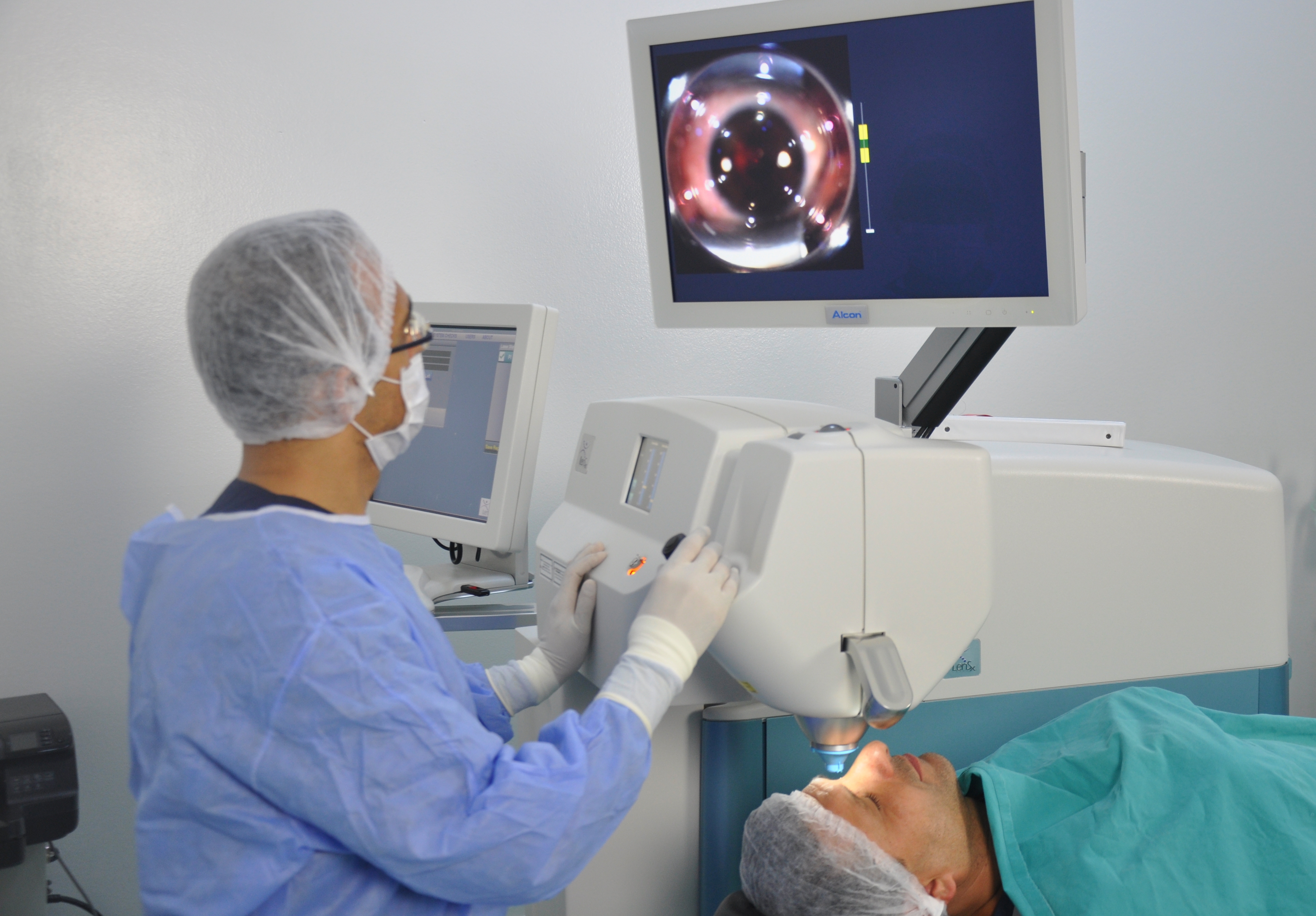
In 2015, Prof Jagat Ram was instrumental in starting state-of-the-art Femtosecond Laser Assisted Cataract Surgery at the PGIMER, Chandigarh and this facility was inaugurated by Union Minister for Health and Family Welfare, J.P Nadda.
If we look back in time, there have been tremendous improvements and refinements in the technique of cataract surgery in children from 1979 till date. The surgical procedure in children for cataract surgery was needling of the congenital and developmental cataract from 1979-1990. Thereafter the surgical procedure advanced to ECCE with IOL implantation from 1985-95. During this period, Prof Ram was instrumental in starting cataract surgery in young children with IOL implantation and performing various complicated cataract surgeries among young children with congenital complications including persistent foetal vessels and other defects for which otherwise no cure was available. He has to his credit surgical innovations for cataract surgery in children.
Prof Ram has contributed significantly in patient care throughout his illustrative career spanning over close to four decades in restoring the vision by modern surgical technique in approximately over 82,000 patients suffering from visual impairment due to cataract. Imbibed with a missionary zeal, he has constantly offered his services in over 135 eye relief services in the rural areas of neighbouring states of Punjab, Haryana and Himachal Pradesh. Prof Ram is a regular participant at the rural camps at Beas Amritsar where extremely poor and deprived individuals are provided with high quality care. He was selected by the Government of India for two years assignment with the Republic of Seychelles as Consultant Ophthalmologist from 2003 to 2005 where he was successful in eliminating the backlog of cataract blindness.
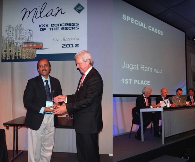
He is recipient of over 30 awards at the International, national and state level including most prestigious International Award named as the Best of the Best Award for Innovation in New Surgical Technique for children with cataract and congenital defects at American Society Cataract and Refractive Society Conference held at San Francisco USA in 2013. He was again awarded the Best of the Best Award at New Orleans USA in 2016. He was awarded Oscar of Paediatric Ophthalmology at the World Congress of Paediatric Ophthalmology and Strabismus at Barcelona in 2015. This achievement was recognized by a number of world leaders in ophthalmology as a tribute to India.
Prof Ram is focused in providing training and mentoring over 250 postgraduate students and ophthalmologists deputed from all over the country and abroad with the most modern surgical technique of phacoemulsification. Prof Ram has delivered over 410 lectures on paediatric and adult cataract in the national and international conferences. As a researcher, he contributed over 300 publications in the National and International Journals including the prestigious New England Journal of Medicine and Lancet. Prof Ram was the only invitee from Asia Pacific region for a special issue on Elimination of Cataract Blindness by Survey of Ophthalmology, which is a unique International honour. Dr Jagat Ram has completed 40 research projects funded by PGI, Government of India and international collaboration. He is on the Editorial Board of Indian Journal of Ophthalmology and as a reviewer for several major International Scientific Journals.
Indian medical community has recognized him as the finest ophthalmic surgeon and a clinician par excellence who provides quality care to all sections of the society. His exceptional surgical qualities and skill have changed lives of several thousand patients especially those belonging to the poor and deprived classes. He is a role model for the medical students and faculty alike.
Contribution as Director of PGIMER, Chandigarh
On March 17, 2017, he was elevated to the post of Director, PGIMER, Chandigarh. After assuming the charge as Director of the Institute, Prof Jagat Ram has initiated the work on the three major projects after approval by the Government of India namely Mother Child Care Centre and Neurosciences Centre at PGI, Chandigarh and setting up the Satellite Centre at Una in Himachal Pradesh. Upgrading these facilities will greatly benefit the patients coming from the neighbouring states.
He is instrumental in negotiations to significantly bring down the cost of orthopaedic implants and cardiac stents within affordable range for common patients. He has also succeeded in channelizing the working of the overcrowded Emergency by close supervision and prompt management by doctors and staff, which has helped in reducing patient’s number and hospital stay.